🧠 Neuroanatomy — Gross Brain
The cerebral cortex is the most superficial layer of the
cerebrum. It and the underlying white matter accounts for the largest
part of the human brain. — Radiopaedia
🎯 Core Concept
The brain has 4 cerebral lobes, each responsible for
different functions. The surface is folded into gyri
(ridges) and sulci (grooves) to maximize surface area
within the skull.
🖼️ Approved Images
Fig 1. Lobes of the Cerebrum
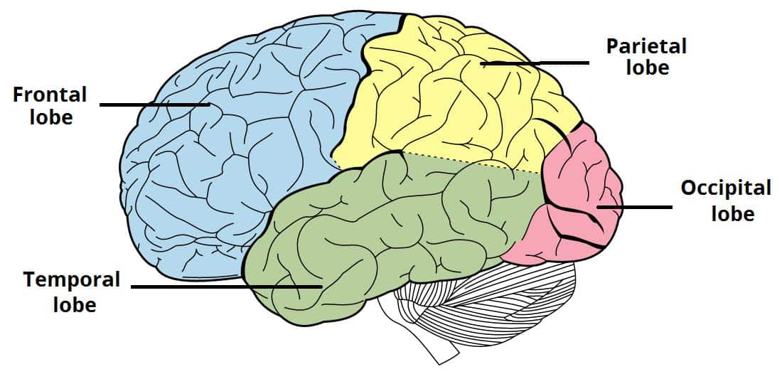 > Source:
TeachMeAnatomy — Neuroanatomy: Structures > Alt:
“Illustration of the lobes of the cerebral cortex, highlighting their
anatomical regions.”
> Source:
TeachMeAnatomy — Neuroanatomy: Structures > Alt:
“Illustration of the lobes of the cerebral cortex, highlighting their
anatomical regions.”
Fig 2. Basal Ganglia
Components
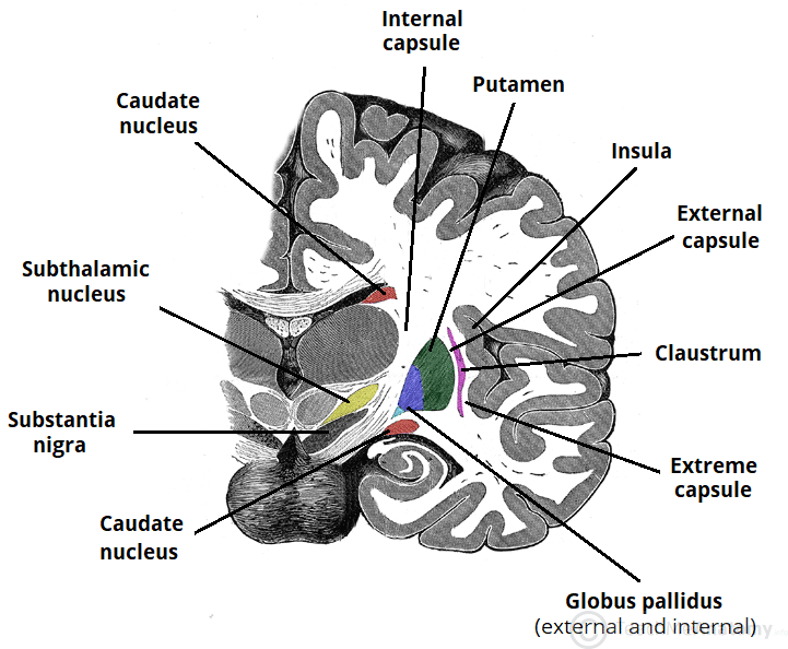 > Source: TeachMeAnatomy —
Neuroanatomy: Structures > Alt: “Illustration of the
components of the basal ganglia and their anatomical relations.”
> Source: TeachMeAnatomy —
Neuroanatomy: Structures > Alt: “Illustration of the
components of the basal ganglia and their anatomical relations.”
🧠 The 4 Cerebral Lobes
| Lobe |
Location |
Key Functions |
Clinical Correlation |
| Frontal |
Anterior to central sulcus, above lateral sulcus |
Motor function, speech (Broca), executive planning, personality,
decision-making, working memory |
Broca aphasia, frontal lobe syndrome, personality changes |
| Parietal |
Behind central sulcus, above lateral sulcus |
Sensory integration, spatial awareness, Wernicke area (language
comprehension), calculation |
Wernicke aphasia, Gerstmann syndrome (angular gyrus lesion) |
| Temporal |
Below lateral sulcus |
Auditory processing, memory (hippocampus), emotion (amygdala),
language (Wernicke) |
Wernicke aphasia, Kluver-Bucy syndrome, memory deficits |
| Occipital |
Posterior to parietal-occipital sulcus |
Primary visual cortex, visual processing, color recognition |
Visual field defects, cortical blindness |
🔑 Key Sulci That Define
Lobe Boundaries
| Sulcus |
Separates |
| Central sulcus (Rolando) |
Frontal ↔︎ Parietal |
| Lateral sulcus (Sylvian) |
Frontal/Parietal ↔︎ Temporal |
| Parieto-occipital sulcus |
Parietal ↔︎ Occipital |
| Calcarine sulcus |
Divides occipital lobe (visual cortex location) |
🧩 Deep Gray Nuclei
(Deep Brain Structures)
These are collections of neurons deep within the
cerebrum, not in the cortex.
The Basal Ganglia Circuit
Cortex → Striatum (Caudate + Putamen) → GPi/SNr → Thalamus → Cortex
              ↑                              ↓
              ←←←←←←←← (Direct/Indirect pathways) ←←←←←←←←
| Structure |
Role |
Clinical Note |
| Caudate nucleus |
Part of striatum; involved in motor planning |
Huntington disease — caudate atrophy |
| Putamen |
Part of striatum; motor, cognitive |
Parkinson — putaminal changes |
| Globus pallidus internus (GPi) |
Output nucleus; inhibits thalamus |
DBS target for Parkinson |
| Subthalamic nucleus (STN) |
Part of indirect pathway |
Lesion → hemiballismus |
| Substantia nigra |
Dopamine production; motor facilitation |
Parkinson — depigmentation |
Other Deep Gray Nuclei
| Structure |
Function |
| Thalamus |
Relay station — sensory, motor, cognitive signals to cortex |
| Amygdala |
Emotion processing, fear response |
| Hippocampus |
Memory consolidation (episodic, spatial) |
🧬 Cerebral Cortex —
Microscopic Structure
Neocortex (6 Layers)
vs Allocortex (3 Layers)
- Neocortex: ~90% of human cortex — 6 layers (I–VI),
responsible for higher functions
- Allocortex: ~10% — 3 layers (hippocampus, olfactory
cortex)
The 6 Layers of Neocortex
| Layer |
Name |
Function |
| I |
Molecular |
Dendritic connections, integration |
| II |
External granular |
Small pyramidal cells, inputs |
| III |
External pyramidal |
Association corticocortical connections |
| IV |
Internal granular |
Thalamic inputs (sensory) |
| V |
Internal pyramidal |
Corticofugal outputs (motor to brainstem/spinal cord) |
| VI |
Multiform |
Outputs to thalamus |
Board Pearl: Layer IV = thalamic input layer —
prominent in primary sensory cortices. Layer V = corticofugal output —
prominent in primary motor cortex (Betz cells).
💡 Gyri — Key Functional
Landmarks
| Gyrus |
Lobe |
Function |
| Precentral gyrus |
Frontal |
Primary motor cortex (M1) — controls voluntary
movement |
| Postcentral gyrus |
Parietal |
Primary somatosensory cortex (S1) — receives
sensory input |
| Superior temporal gyrus |
Temporal |
Wernicke area (language comprehension) — usually LEFT
hemisphere |
| Inferior frontal gyrus |
Frontal |
Broca area — speech production — usually LEFT hemisphere |
| Angular gyrus |
Parietal |
Language, calculation, conceptual processing |
| Parahippocampal gyrus |
Temporal |
Memory encoding |
🔬 Surface Area — Why Folding
Matters
- The cerebral cortex contains ~16 billion
neurons
- Two-thirds of the cortex lies within sulci (hidden
in the grooves)
- One-third is exposed on gyri (visible on the
surface)
- This folding allows ~2,200 cm² of cortical surface to fit inside the
skull
📋 Clinical Pearls
| Scenario |
Lesion Localization |
| Expressive (non-fluent) aphasia with intact comprehension |
Broca area — left inferior frontal gyrus |
| Receptive (fluent) aphasia with poor comprehension |
Wernicke area — left superior temporal gyrus |
| Inability to recognize objects (visual) |
Occipital lobe (inferior visual association
cortex) |
| Personality changes + disinhibition |
Frontal lobe (especially orbitofrontal) |
| Loss of sensation on one side of body |
Postcentral gyrus (contralateral) |
| Weakness of one side of body |
Precentral gyrus (contralateral) |
🎯 Brodmann Areas — High Yield
| Area |
Location |
Function |
| Brodmann 4 |
Precentral gyrus |
Primary motor cortex |
| Brodmann 3,1,2 |
Postcentral gyrus |
Primary somatosensory cortex |
| Brodmann 44,45 |
Inferior frontal gyrus |
Broca area (speech production) |
| Brodmann 22 |
Superior temporal gyrus |
Wernicke area (speech comprehension) |
| Brodmann 17 |
Calcarine sulcus (occipital) |
Primary visual cortex |
| Brodmann 41,42 |
Transverse temporal gyrus |
Primary auditory cortex |
🗺️ Functional Organization
ANTERIOR (Frontal)          →          →          →    POSTERIOR (Occipital)

Prefrontal    Motor        Sensory       Parietal      Visual
Cortex        Cortex       Cortex        Association   Cortex
(Executive)    (M1)         (S1)          Cortex        (V1)
📚 Sources
- Alberstone et al. — Anatomic Basis of
Neurologic Diagnosis (2nd ed, 2023)
- Bradley — Neurology in Clinical Practice
(8th ed)
- Adams & Victor — Principles of
Neurology (12th ed, 2023)
- Radiopaedia — Cerebral
cortex
- TeachMeAnatomy — Neuroanatomy:
Structures
- [[wiki/foundation/NEUROANATOMY_BLOOD_SUPPLY|Blood Supply — Cerebral
Circulation]]
- [[wiki/foundation/NEUROANATOMY_LIMBIC|Limbic System — Memory &
Emotion]]
- [[wiki/foundation/NEUROANATOMY_BASAL_GANGLIA|Basal Ganglia — Motor
Circuits]]
- [[wiki/foundation/CRANIAL_NERVES|Cranial Nerves Overview]]
🃏 Anki Prompts
| Front |
Back |
| What separates the frontal from parietal lobe? |
Central sulcus (Rolando) |
| What separates the frontal from temporal lobe? |
Lateral sulcus (Sylvian) |
| Broca area is in which gyrus and lobe? |
Inferior frontal gyrus, Frontal lobe |
| Wernicke area is in which gyrus and lobe? |
Superior temporal gyrus, Temporal lobe |
| What are the 4 cerebral lobes? |
Frontal, Parietal, Temporal, Occipital |
| What are the components of the basal ganglia? |
Caudate, Putamen, Globus pallidus, Subthalamic nucleus, Substantia
nigra |
| Which layer of neocortex receives thalamic input? |
Layer IV (internal granular) |
| Which layer contains Betz cells (corticofugal output)? |
Layer V (internal pyramidal) |
| Lesion of the dominant hemisphere angular gyrus causes? |
Gerstmann syndrome (agraphia, acalculia, finger agnosia, L-R
confusion) |
Last updated: 2026-04-24 | Phase 1 — Foundation | Status: 🔄 In
Progress